
1. Introduction to Ti Sapphire Crystal
1.1 History of Ti Sapphire Crystal
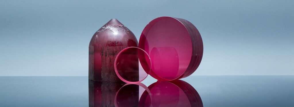
Ti Sapphire crystal was first discovered in the 1960s by a team of researchers led by Robert L. Byer at Stanford University. The crystal is created by doping Sapphire, a crystalline form of aluminum oxide, with small amounts of titanium ions. The resulting crystal exhibits unique optical and physical properties, making it ideal for various medical applications.
1.2 Physical and Optical Properties
Ti:Sapphire crystal has several unique physical and optical properties, making it highly desirable for medical applications. The crystal has a broad absorption band in the visible region of the spectrum, making it highly efficient at absorbing light. It also has a broad emission band, enabling ultrashort laser pulses in the femtosecond range.
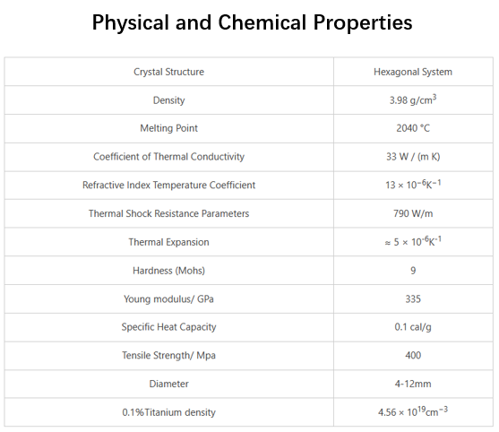
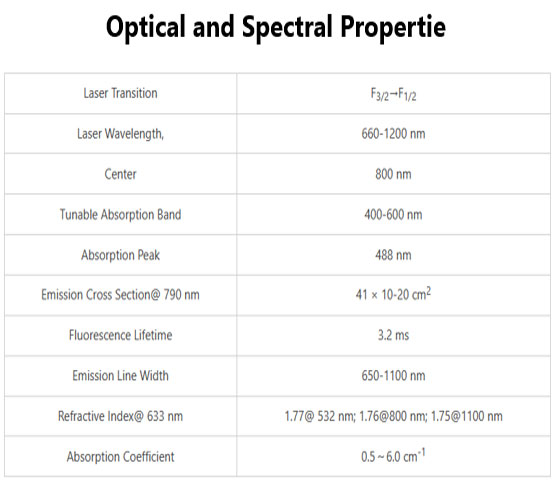
The crystal has a high gain, low lasing threshold, and broad bandwidth, making it an ideal candidate for various medical applications. The crystal is highly transparent in the near-infrared region of the spectrum, which is ideal for medical imaging and phototherapy applications.
1.3 Importance in Medical Applications
The unique properties of Ti Sapphire crystal have made it an important material for various medical applications. The crystal can be used in medical imaging, photodynamic therapy, laser surgery, tissue engineering, and gene therapy.
In medical imaging, Ti:Sapphire crystal is used in various techniques such as optical coherence tomography, multiphoton microscopy, and second-harmonic generation imaging. The crystal’s ability to generate ultrashort laser pulses in the femtosecond range makes it ideal for photodynamic therapy and laser surgery. Ti Sapphire crystal can also be used in tissue engineering to stimulate cell growth and differentiation.
1.4 Current State of Research
Significant research has been conducted on the use of Ti:Sapphire crystal in medical applications, with many promising results. In recent years, a particular focus has been on developing new techniques for using Ti Sapphire crystal in gene therapy.
Despite the promising results, significant challenges must be overcome before Ti Sapphire crystal can be widely used in medical applications. These challenges include regulatory hurdles, cost considerations, and the need for further research and development.
1.5 Conclusion
In conclusion, Ti:Sapphire crystal is a highly versatile material with unique physical and optical properties that make it ideal for various medical applications. Its importance in medical applications will likely grow as new techniques and applications are developed. Further research is needed to fully understand the potential of Ti Sapphire crystal in medical applications, but the outlook is highly promising.
2. Ti Sapphire Crystal Synthesis and Characterization
2.1 Synthesis of Ti Sapphire Crystal
Ti:Sapphire crystal can be synthesized using various methods, including the Czochralski, flux, and hydrothermal methods. The Czochralski method involves slowly pulling a seed crystal from a melt of Sapphire and titanium oxide. In contrast, the flux method involves dissolving Sapphire and titanium oxide in a flux material and slowly cooling the resulting solution to allow the crystal to form. The hydrothermal method involves placing a mixture of sapphire and titanium oxide in a high-pressure chamber filled with water and heating the chamber to allow the crystal to form.
2.2 Characterization of Ti Sapphire Crystal
Characterization of Ti:Sapphire crystal is an important step in understanding its properties and applications. Various techniques are used to characterize Ti Sapphire crystals, including X-ray diffraction, Raman spectroscopy, and scanning electron microscopy.
X-ray diffraction is a powerful technique for determining the crystal structure of Ti Sapphire crystals. This technique involves shining X-rays onto the crystal and measuring the diffraction pattern produced. This pattern can be used to determine the crystal structure and quality of the crystal.
Raman spectroscopy is another technique used to characterize Ti:Sapphire crystals. This technique involves a laser on the crystal and measuring the resulting scattered light. The scattered light provides information about the crystal’s vibrational modes, which can be used to determine its properties.
Scanning electron microscopy is a technique used to visualize the surface of Ti:Sapphire crystals. This technique involves scanning the surface of the crystal with a beam of electrons and measuring the resulting signal. This signal can create an image of the crystal’s surface, providing information about its quality and structure.
2.3 Advances in Ti Sapphire Crystal Synthesis and Characterization
Significant advances have been made in synthesizing and characterizing Ti:Sapphire crystals in recent years. Researchers have developed new methods for synthesizing Ti Sapphire crystals, such as using microwaves to improve the crystal’s quality and using laser-induced nucleation to control crystal growth.
Advances in characterization techniques have also enabled researchers to understand Ti:Sapphire crystals’ properties better. For example, researchers have used Raman spectroscopy to study the effects of doping on the crystal’s properties and X-ray diffraction to study the crystal’s structural properties.
2.4 Conclusion
In conclusion, synthesizing and characterizing Ti:Sapphire crystals are crucial to understanding their properties and potential medical applications. Advances in synthesis and characterization techniques have enabled researchers to understand the properties of Ti Sapphire crystals better and develop new methods for their synthesis. Further research in this area is needed to fully exploit the potential of Ti Sapphire crystals in medical applications.
3. Ti Sapphire Crystal for Medical Imaging
3.1 Introduction
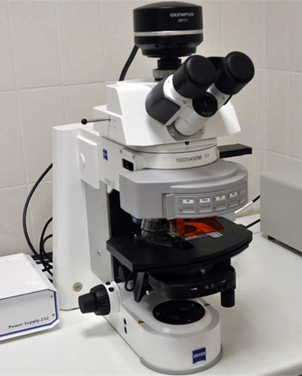
Ti:Sapphire crystal is used in various medical imaging techniques, including optical coherence tomography (OCT), multiphoton microscopy, and second-harmonic generation imaging. In this chapter, we will discuss the principles of these imaging techniques and Ti Sapphire crystal’s role in each of them.
3.2 Optical Coherence Tomography
Optical coherence tomography (OCT) is a non-invasive imaging technique that uses low-coherence light to create high-resolution images of biological tissues. Ti Sapphire crystals are used in OCT as a light source due to their ability to generate ultrashort laser pulses in the femtosecond range.
In OCT, the light generated by the Ti Sapphire crystal is split into a sample arm and a reference arm. The light in the sample arm is directed at the biological tissue being imaged, while the light in the reference arm is directed at a mirror. The light reflected from the biological tissue and the mirror is then combined and analyzed to create an image of the tissue.
Ti Sapphire crystals are used in OCT because they generate short, high-intensity laser pulses, which can penetrate deep into biological tissues. This allows for high-resolution images to be obtained from deep within tissues.
3.3 Multiphoton Microscopy
Multiphoton microscopy is a technique that uses ultrashort laser pulses to image biological tissues. The technique involves using a Ti Sapphire crystal to generate femtosecond laser pulses focused on the biological tissue being imaged.
When the laser pulses interact with the biological tissue, they cause two or more photons to be absorbed simultaneously, which results in fluorescence emission. The emitted fluorescence is detected and used to create an image of the tissue.
Ti Sapphire crystals are used in multiphoton microscopy because they generate ultrashort laser pulses in the femtosecond range, which are required for this imaging technique. The high peak power of the laser pulses also allows for deeper penetration into tissues, which results in higher-resolution images.
3.4 Second-Harmonic Generation Imaging
Second-harmonic generation imaging is a technique that uses ultrashort laser pulses to image biological tissues. The technique involves using a Ti Sapphire crystal to generate femtosecond laser pulses focused on the biological tissue being imaged.
When the laser pulses interact with the biological tissue, they cause second-harmonic generation, which is the emission of photons with twice the incident laser frequency. The emitted photons are detected and used to create an image of the tissue.
Ti Sapphire crystals are used in second-harmonic generation imaging due to their ability to generate ultrashort laser pulses in the femtosecond range. The high peak power of the laser pulses also allows for deeper penetration into tissues, which results in higher-resolution images.
3.5 Advantages and Limitations
Using Ti Sapphire crystals in medical imaging has several advantages, including generating ultrashort laser pulses, which can penetrate deep into biological tissues, resulting in high-resolution images. Ti Sapphire crystals are also highly efficient and have a broad bandwidth, which allows for a wide range of imaging applications.
However, there are also limitations to the use of Ti:Sapphire crystals in medical imaging. These limitations include the need for expensive equipment and skilled operators and the potential for tissue damage due to the high intensity of the laser pulses.
3.6 Conclusion
In conclusion, Ti Sapphire crystals are important components of several medical imaging techniques, including OCT, multiphoton microscopy, and second-harmonic generation imaging. The unique properties of Ti Sapphire crystals, such as their ability to generate ultrashort laser pulses, make them ideal for medical imaging applications. Further research is needed to fully understand the potential of Ti Sapphire crystals in medical imaging and to overcome the limitations associated with their use. Nevertheless, the outlook for using Ti Sapphire crystals in medical imaging is highly promising.
4. Ti Sapphire Crystal for Phototherapy
4.1 Introduction
Ti Sapphire crystal has emerged as a promising material for phototherapy applications, including photodynamic therapy (PDT) and laser therapy. In this chapter, we will discuss the principles of phototherapy and Ti Sapphire crystal’s role in these applications.
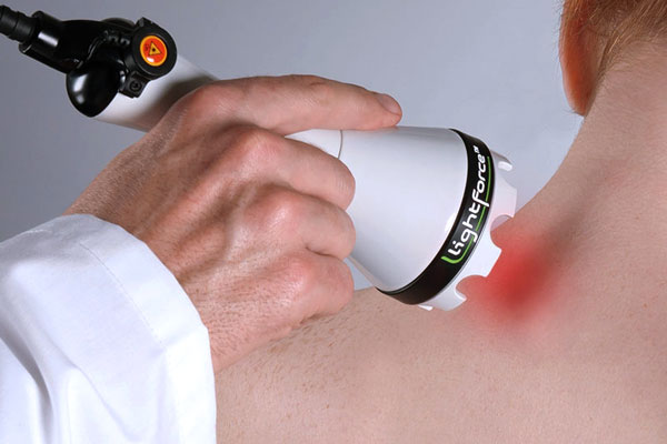
4.2 Photodynamic therapy
Photodynamic therapy (PDT) is a non-invasive treatment that uses photosensitizing agents and light to treat various medical conditions, including cancer, macular degeneration, and acne. The photosensitizing agent is administered to the patient and is taken up by the target cells. The Ti Sapphire crystal is a light source to activate the photosensitizer and triggers the therapeutic response.
When the photosensitizer is exposed to light from the Ti Sapphire crystal, it absorbs the light energy. It undergoes a series of reactions that produce reactive oxygen species (ROS), such as singlet oxygen. These ROS then damage the target cells, leading to their destruction.
Ti Sapphire crystals are ideal for PDT applications because they generate high-intensity laser pulses in the femtosecond range, which can activate the photosensitizer with high precision. Ti Sapphire crystals also allow for deeper penetration of the light into the tissue, which increases the effectiveness of the treatment.
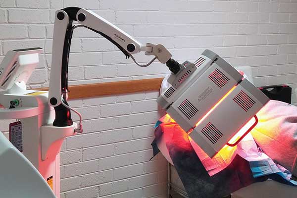
4.3 Laser Therapy
Laser therapy is a non-invasive treatment involving laser light to treat various medical conditions, including dermatological conditions, musculoskeletal disorders, and pain. Ti Sapphire crystals are the light source in laser therapy because they generate high-intensity laser pulses in the femtosecond range.
The laser light generated by the Ti Sapphire crystal is absorbed by the target tissue, causing various physiological responses, including increased blood flow, tissue regeneration, and pain relief. The use of Ti Sapphire crystals allows for precise targeting of the tissue and increased effectiveness of the treatment.
4.4 Advantages and Limitations
The use of Ti Sapphire crystals in phototherapy has several advantages, including their ability to generate high-intensity laser pulses in the femtosecond range, which allows for precise tissue targeting and increased treatment effectiveness. Ti Sapphire crystals are also highly efficient and have a broad bandwidth, which allows for a wide range of phototherapy applications.
However, there are also limitations to the use of Ti Sapphire crystals in phototherapy. These limitations include the potential for tissue damage due to the high intensity of the laser pulses and the need for expensive equipment and skilled operators.
4.5 Conclusion
In conclusion, Ti Sapphire crystals have emerged as promising materials for phototherapy applications, including PDT and laser therapy. The unique properties of Ti:Sapphire crystals, such as their ability to generate high-intensity laser pulses in the femtosecond range, make them ideal for phototherapy applications. Further research is needed to fully understand Ti:Sapphire crystals’ potential in phototherapy and overcome the limitations associated with their use. Nevertheless, the outlook for using Ti Sapphire crystals in phototherapy is highly promising.

Frank
Frank graduated from the University of Shanghai for Science and Technology, majoring in optics. As a technical engineer at Crylink Company, he deeply understands crystal materials and laser components.
Related Video(s) with this Article
Related Product(s) with this Article
Related Application(s) with this Article